Students can Download Bio Zoology Chapter 7 Body Fluids and Circulation Questions and Answers, Notes Pdf, Samacheer Kalvi 11th Bio Zoology Book Solutions Guide Pdf helps you to revise the complete Tamilnadu State Board New Syllabus and score more marks in your examinations.
Tamilnadu Samacheer Kalvi 11th Bio Zoology Solutions Chapter 7 Body Fluids and Circulation
Samacheer Kalvi 11th Bio Zoology Body Fluids and Circulation Text Book Back Questions and Answers
I. Multiple Choice Questions
Question 1.
What is the function of lymph?
(a) Transport of O2 into brain
(b) Transport of CO2 into lungs
(c) Bring interstitial fluid in blood
(d) Bring RBC and WBC in lymph node
Answer:
(c) Bring interstitial fluid in blood
Question 2.
Which one of the following plasma proteins is involved in the coagulation of blood?
(a) Globulin
(b) Fibrinogen
(c) Albumin
(d) Serum amylase
Answer:
(b) Fibrinogen
![]()
Question 3.
Which of the following WBCs are found in more numbers?
(a) Eosinophil
(b) Neutrophil
(c) Basophil
(d) Monocyte
Answer:
(b) Neutrophil
Question 4.
Which of the following is not involved in blood clotting?
(a) Fibrin
(b) Calcium
(c) Platelets
(d) Bilirubin
Answer:
(d) Bilirubin
![]()
Question 5.
Lymph is colourless because ………….
(a) WBC are absent
(b) WBC are present
(c) Haemoglobin is absent
(d) RBC are absent
Answer:
(c) Haemoglobin is absent
Question 6.
Blood group is due to the presence or absence of surface
(a) Antigens on the surface of WBC
(b) Antibodies on the surface of RBC
(c) Antigens on the surface of RBC
(d) Antibodies on the surface of WBC
Answer:
(c) Antigens on the surface of RBC
![]()
Question 7.
A person having both antigen A and antigen B on the surface of RBCs belongs to blood group
(a) A
(b) B
(c) AB
(d) O
Answer:
(c) AB
Question 8.
Erythroblastosis foetalis is due to the destruction of …………..
(a) Foetal RBCs
(b) Foetus suffers from atherosclerosis
(c) Foetal WBCs
(d) Foetus suffers from mianmata
Answer:
(a) Foetal RBCs
Question 9.
Dub sound of heart is caused by
(a) Closure of atrio-ventricular valves
(b) Opening of semi-lunar valves
(c) Closure of semi-lunar valves
(d) Opening of atrio-ventricular valves
Answer:
(c) Closure of semi-lunar values
![]()
Question 10.
Why is the velocity of blood flow the lowest in the capillaries?
(a) The systemic capillaries are supplied by the left ventricle, which has a lower cardiac output than the right ventricle.
(b) Capillaries are far from the heart, and blood flow slows as distance from the heart increases.
(c) The total surface area of the capillaries is larger than the total surface area of the arterioles.
(d) The capillary walls are not thin enough to- allow oxygen to exchange with the cells.
(e) The diastolic blood pressure is too low to deliver blood to the capillaries at a high flow rate.
Answer:
(c) The total surface area of the capillaries is larger than the total surface area of the arterioles.
Question 11.
An unconscious patient is rushed into the emergency room and needs a fast blood transfusion. Because there is no time to check her medical history or determine her blood type, which type of blood should you as her doctor, give her?
(a) A+
(b) AB
(c) O+
(d) O–
Answer:
(c) O+
![]()
Question 12.
Which of these functions could or could not be carried out by a red blood cell?
(a) Protein synthesis
(b) Cell division
(c) Lipid synthesis
(d) Active transport
Answer:
(a) Protein synthesis: RBCs do not have ribosomes which are important for protein synthesis, They are concerned with transport of respiratory gases alone. Hence protein synthesis
could not take place in RBCs.
(b) Cells division: RBCs do not have numbers. They are produced in the bone marrow. They do not involve in cell division.
(c) Lipid Synthesis: Lipid synthesis occurs in endoplasmic reticulum (ER) and golgi complex. The ER is absent in RBCs. Hence lipid synthesis does not take place in RBCs.
(d) Active transport: Transport of respiratory gases between the alveoli to the blood vessels, blood vessel to the cells and vice versa take place due to difference in the partial pressure of O2 and CO2., Active transport of materials against concentration gradient does not take place in RBCs.
Question 13.
At the venous end of the capillary bed, the osmotic pressure is …………….
(a) Greater than the hydrostatic pressure
(b) Result in net outflow of fluids
(c) Results in net absorption of fluids
(d) No change occurs
Answer:
(a) Greater than the hydrostatic pressure
![]()
Question 14.
A patient’s chart reveals that he has a cardiac output of 7500mL per minute and a stroke volume of 50 mL. What is his pulse rate (in beats / min)
(a) 50
(b) 100
(c) 150
(d) 400
Answer:
(c) 150
Question 15.
At any given time there is more blood in the venous system than that of the arterial system. Which of the following features of the veins allows this?
(a) relative lack of smooth muscles
(b) presence of valves
(c) proximity of the veins to lymphatic’s
(d) thin endothelial lining
Answer:
(a) relative lack of smooth muscles
II. Short Answer Questions
Question 16.
Distinguish between arteries and veins?
Answer:
| Arteries | Veins |
| 1. Arteries are the blood vessels that carry blood away from the heart. | 1. Veins are the blood vessels that carry blood to the heart. |
| 2. Arteries carry oxygenated blood except pulmonary artery. | 2. Veins carry deoxygenated blood except pulmonary veins. |
| 3. Arteries usually lie deep inside the body. | 3. Veins are usually located superficially. |
| 4. These are thick walled. | 4. These are thin walled. |
| 5. These do not have valves. | 5. These have semilunar valves. |
| 6. Blood pressure is high. | 6. Blood pressure is low. |
![]()
Question 17.
Distinguish between open and closed circulation?
Answer:
| Open circulation | Closed circulation |
| 1. Open circulation, haemolymph is pumped by the heart which flows through blood vessels into the haemocoel. | 1.In closed circulation, blood is pumped by the heart and flows through blood vessels |
| 2. It is seen in arthropods and most molluscs. | 2. It is seen in annelids, cephalopods and vertebrates |
Question 18.
Distinguish between mitral valve and semi lunar valve?
Answer:
| Mitral valve |
Semilunar vales |
| 1. The valve present between the left atrium left ventricle is called mitral valve. | 1. The valves present at the openings of right and left ventricles into the pulmonary artery and aorta are semilunar valves. |
| 2. It is made of two flaps. | 2. These are of three half moon shaped cusps. |
![]()
Question 19.
Right ventricular wall is thinner than the left ventricular wall. Why?
Answer:
The wall of the left ventricle is thick
Reason:
- As the right ventricle constricts the deoxygenated blood through the pulmonary vein goes to the lungs only. These may not be necessary for much blood pressure for this.
- As the left ventricle constricts through the dorsal aorta blood goes to all the parts of the body.
- For this action, more pressure is needed. Hence the wall of the left ventricle is thick. It gives more pressure on the contraction of the ventricle to distribute the blood.
Question 20.
What might be the effect on a person whose diet has less iron content?
Answer:
A person whose diet has less iron content will become anaemic. The haemoglobin content of the blood will be less. The volume of oxygen carried by RBCs gets reduced. He/she may experience tiredness, weakness, fatigue etc. In order to overcome this deficiency one has to take iron-rich diet.
![]()
Question 21.
Describe the mechanism by which the human heartbeat is initiated and controlled?
Answer:
The rhythmic contraction and expansion of heart is called heartbeat. The contraction of the heart is called systole and the relaxation of the heart is called diastole. The human heart is myogenic. The pacemaker cells are located in the right sinoatrial (SA) node.
On the left side of the right atrium, there is a mode called auriculo ventricular node (AV). Two special cardiac muscle fibers which originate from the AV node are called the bundle of His. It runs down into the interventricular spectrum and the fibres spread into the ventricle as the Purkinje fibres.
The pacemaker cells produce excitation through depolarization of their cell membrane. Early depolarization is slow and takes place by sodium influx and reduction in potassium efflux. Minimum potential is required to activate voltage gated calcium (Ca+) channels that cause rapid depolarization which results in action potential. The pace maker cells repolarise slowly via K+ efflux.
![]()
Question 22.
What is lymph? Write its function?
Answer:
The fluid inside the lymphatics is called lymph. Uses:
- It helps in transporting nutrients hormones oxygen within the body cells.
- It keeps the body cells moist.
- The lymph nodes synthesize lymphocytes.
- It keeps the blood volume uniformly.
- Fats are absorbed through lymph in the lacteals present in the villi of the intestinal wall.
![]()
Question 23.
What are the heart sounds? When and how are these sounds produced?
Answer:
During each cardiac cycle due to the action of valves, two sounds like lub and dub are produced. These sounds are known as cardiac sounds.
- Lub sound: During the contraction of the ventricle, the closure of tricuspid and bicuspid valves make this sound.
- Dub sound: During the relaxation of the ventricle, the valves of the pulmonary artery and dorsal aorta close and make this sound.
Question 24.
Select the correct biological term. Lymphocytes, red cells, leukocytes, plasma, erythrocytes, white cells, haemoglobin, phagocyte, platelets, blood clot?
Question (a)
Disc-shaped cells which are concave on both sides?
Answer:
Red blood cells
Question (b)
Most of these have a large, bilobed nucleus?
Answer:
Leucocytes
Question (c)
Enable red cells to transport blood?
Answer:
Hemoglobin
Question (d)
The liquid part of the blood?
Answer:
plasma
Question (e)
Most of them move and change shape like an amoeba?
Answer:
phagocyte
Question (f)
Consists of water and important dissolved substances?
Answer:
plasma
Question (g)
Destroyed in the liver and spleen after circulating in the blood for four months?
Answer:
RBCs
Question (h)
The substances which give red colour to their cells?
Answer:
haemoglobin
Question (i)
Another name for red blood cells?
Answer:
Erythrocytes
Question (j)
Blood that has been changed to a jelly?
Answer:
Blood clot
Question (k)
A word that me cell eater?
Answer:
Phagocyte
Question (l)
Cells without a nucleus?
Answer:
Red blood cells
Question (m)
White cells made in the lymphatic tissue?
Answer:
Lymphocytes
Question (n)
Blocks wound and prevent excessive bleeding?
Answer:
Platelets
Question (o)
Fragment of cells which are made in the bone marrow?
Answer:
Erythrocytes
Question (p)
Another name for white blood cells?
Answer:
Leucocytes
Question (q)
Slowly releases oxygen to blood cells?
Answer:
Red cells
Question (r)
Their function is to help blood clot in wounds?
Answer:
Platelets
![]()
Question 25.
Select the correct biological term?
Answer:
Cardiac muscle, atria, tricuspid valve, systole, auricles, arteries, diastole, ventricles, bicuspid valve, pulmonary artery, cardiac cycle, semilunar valve, veins, pulmonary vein, capillaries, vena cava, aorta?
Question (a)
The main artery of the blood?
Answer:
Aorta
Question (b)
Valves between the left atrium and ventricle?
Answer:
Bicuspid valve
Question (c)
The technical name for relaxation of the heart?
Answer:
Diastole
Question (d)
Another name for atria?
Answer:
Arteries
Question (e)
The main vein?
Answer:
Vena cava
Question (f)
Vessels which carry blood away from the heart?
Answer:
Arteries
Question (g)
Two names for the upper chambers of the heart?
Answer:
Atria
Question (h)
Thick-walled chambers of the heart?
Answer:
Atria
Question (i)
Carries blood from the heart to the lungs?
Answer:
Pulmonary Artery
Question (j)
Takes about 0.8 sec to complete?
Answer:
Cardiac cycle
Question (k)
Valves situated at the point where blood flows out of the heart?
Answer:
Semilunar valves
Question (l)
Vessels which carry blood towards the heart?
Answer:
Veins
Question (m)
Carries blood from the lungs to the heart?
Answer:
Pulmonary veins
Question (n)
The two lower chambers of the heart?
Answer:
Ventricles
Question (o)
Prevent blood from re-entering the ventricles after entering the aorta?
Answer:
Semilunar valves
Question (p)
The technical name for one heartbeat?
Answer:
Cardiac cycle
Question (q)
Valves between right atrium and ventricles?
Answer:
Tricuspid valve
Question (r)
The technical name for the contraction of the heart?
Answer:
Systole
Question (s)
Very narrow blood vessels?
Answer:
Capillaries
![]()
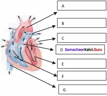
Question 26.
Name and label the given diagram to show A, B, C, D, E, F, and G?
(A) Aorta
(B) Pulmonary trunk
(C) Left pulmonary veins
(D) Blocking the action of vasoconstrictor lowers the blood pressure. Give reasons.
(E) What is the role of ACH inhibitors in reducing blood pressure?
(F) What conditions one might expect if the blood pressure is not controlled?
Answer:
(A) Aortic arch
(B) Left pulmonary artery
(C) Left pulmonary veins
(D) Pulmonary trunk
(E) Left ventricle
(F) Right ventricle
(G) Inferior vena cava
Samacheer Kalvi 11th Bio Zoology Body Fluids and Circulation Additional Questions & Answers
I. Multiple Choice Questions
Choose The Correct Answer
Question 1.
Which of the following is not the function of the circulatory system?
(a) Transport of respiratory gases
(b) Carrying of digested food materials
(c) Transport of hormones to the target organism
(d) Removal of nitrogenous wastes from the body
Answer:
(d) Removal of nitrogenous wastes from the body
![]()
Question 2.
Which is known as a liquid connective tissue?
(a) Plasma
(b) Blood
(c) Serum
(d) lymph
Answer:
(b) blood
Question 3.
What is the function of albumin?
(a) Transport of hormones
(b) Blood clothing
(c) Maintenance of osmotic pressure
(d) Immunity
Answer:
(c) Maintenance of osmotic pressure
Question 4.
Fibrinogen is concerned with ………….
(a) Transport of ions
(b) Transport of liquids
(c) Transport of hormones
(d) Coagulation of blood
Answer:
(d) Coagulation of blood
![]()
Question 5.
The red colour of the RBC is due to the presence of a respiratory pigment ……………
(a) Haemoerythrin
(b) Haemoglobin
(c) Haemocyanin
(d) Chlorocronin
Answer:
(b) Haemoglobin
Question 6.
Which of the following are non-nucleated cells?
(a) WBCs
(b) Nerve cell
(c) RBCs
(d) Muscle cell
Answer:
(c) RBCs
Question 7.
What is hematocrit/packed cells volume?
(a) The ratio of WBCs to blood plasma
(b) The ratio of RBCs to blood plasma
(c) The ratio of platelets to blood plasma
(d) The ratio of plasma and blood cells
Answer:
(b) The ratio of RBCs to blood plasma
![]()
Question 8.
Which of the following is abundant in blood?
(a) Neutrophils
(b) Eosinophils
(c) Basophils
(d) Lymphocytes
Answer:
(a) Neutrophils
Question 9.
…………… are the blood cells which have two lobes which are joined by thin strands.
(a) Neutrophils
(b) Basophils
(c) Eosinophils
(d) Lymphocytes
Answer:
(c) Eosinophils
![]()
Question 10.
What is the percentage of lymphocytes among WBCs?
(a) 0.5 to 1.0%
(b) 1-3%
(c) 65%
(d) 28%
Answer:
(d) 28%
Question 11.
The macrophages in the sinusoids of the liver are called ………….
(a) Microglia
(b) Kupffer cells
(c) Alveolar macrophages
(d) Lymphocytes
Answer:
(b) Kupffer cells
Question 12.
‘A’ blood group has ………………….. antigen and …………………. antibody
(a) A anti B
(b) AB, no antibodies
(c) No antigen, anti-A, Anti B
(d) B, Anti A
Answer:
(d) B, Anti A
![]()
Question 13.
Erythroblastosis foetalis is a condition of incompatibility related to ……………..
(a) Rh antigen and Rh antibodies
(b) Anti A and antigen B
(c) Anti B and antigen A
(d) Antigens A and B
Answer:
(a) Rh antigen and Rh antibodies
Question 14.
The conversion of prothrombin into thrombin occurs in the presence of ……………..
(a) Potassium and vitamin D
(b) Sodium and vitamin B,2
(c) Calcium and vitamin K
(d) Iodine and vitamin E
Answer:
(c) Calcium and vitamin K
Question 15.
………………….. is the exceptional artery that carries deoxygenated blood.
(a) Pulmonary artery
(b) Carotid artery
(c) Coronary artery
(d) Femoral artery
Answer:
(a) Pulmonary artery
![]()
Question 16.
Pulmonary veins carry ………………. blood from lungs to ……………….
(a) Oxygenated, right auricle
(b) Deoxygenated, right auricle
(c) Deoxygenated, left auricle
(d) Oxygenated, left auricle
Answer:
(c) Deoxygenated, left auricle
Question 17.
The blood vessels that supply blood to the cardiac muscles with all nutrients are
(a) Coronary arteries
(b) Cerebral arteries
(c) Aorta
(d) Pulmonary veins
Answer:
(a) Coronary arteries
Question 18.
The opening between the left atrium and left ventricle is guarded by …………….
(a) Semilunar valves
(b) Mitral valve
(c) Tricuspid valve
(d) Flaps
Answer:
(b) Mitral valve
Question 19.
The heart normally beats times …………………. per minute in a human adult.
(a) 60-62
(b) 50-52
(c) 70-72
(d) 90-92
Answer:
(c) 70-72
![]()
Question 20.
Which wave shape occurs from the start of depolarisation of the atria to the beginning of ventricular depolarisation?
(a) P wave
(b) ST segment
(c) QRS complex
(d) PQ interval
Answer:
(d) PQ interval
Question 21.
In the systemic circulation, blood from the …………………. ventricle is carried by a network of arteries, arterioles, and capillaries to the tissues.
(a) Deoxygenated right
(b) Oxygenated left
(c) Oxygenated right
(d) Deoxygenated left
Answer:
(b) Oxygenated, left
![]()
Question 22.
Which hormone increases the heartbeat?
(a) Acetylcholine
(b) Gastrin
(d) Epinephrine
(d) Oxytocin
Answer:
(c) Epinephrine
Question 23.
Thrombus in a coronary artery results in ………….
(a) Heart attack
(b) Stroke
(c) Hypertension
(d) Heart failure
Answer:
(a) Heart attack
Question 24.
Cerebral infarction is called ……….
(a) Heart attack
(b) Hypertension
(c) Heart failure
(d) Stroke
Answer:
(d) Stroke
![]()
Question 25.
The failure of the heart to pump out the normal stroke volume is a condition called ………….
(a) Cerebral thrombosis
(b) Hypertension
(c) Myocardial infarction
(d) Rheumatoid heart disease
Answer:
(c) Myocardial infarction
Question 26.
In which condition the heart muscles do not get oxygen supply?
(a) Stroke
(b) Ischemic heart disease
(c) Hypertension
(d) Heart attack
Answer:
(b) Ischemic heart disease
Question 27.
Which of the following is the autoimmune disease that damages the heart?
(a) Ischemic heart disease
(b) Myocardial infarction
(c) Cerebral thrombosis
(d) Rheumatic fever
Answer:
(d) Rheumatic fever
Question 28.
The life saving procedure, CPR was first used by ……………
(a) William Harvey
(b) Carl Landsteiner
(c) James Elam and Peter Safar
(d) Raymond de Viessens
Answer:
(c) James Elam and Peter Safar
![]()
II. Fill in the Blanks
Question 1.
The tissue fluid that surrounds the cell is ………..
Answer:
Interstitial fluid.
Question 2.
The fluid component of the blood is …………
Answer:
Plasma
Question 3.
The blood flowing into the capillary from an arteriole has a high …………. pressure.
Answer:
Hydrostatic
![]()
Question 4.
………….. is the plasma protein that facilitates the transport of ions, hormones, lipids, and assists in immune function.
Answer:
Globulin
Question 5.
………….. is the respiratory pigment that facilitates the transport of gases.
Answer:
Haemoglobin
Question 6.
The RBCs are destroyed in the liver and ………….
Answer:
Spleen
Question 7.
………….. is the hormone that helps in the differentiation of stem cells of bone marrow into erythrocytes.
Answer:
Erythropoietin
![]()
Question 8.
The ratio of red blood cells to blood plasma is expressed is …………
Answer:
Haematocrit
Question 9.
The granulocytes have in the cytoplasm.
Answer:
Granules
Question 10.
Neutrophils are also called …………
Answer:
Heterophils/polymorphonuclear cells
Question 11.
…………… have distinctly dilobed nucleus and the lobes are joined by thin strands.
Answer:
Eosinophils
Question 12.
Basophils secrete substances such as ……………… serotonin and histones.
Answer:
Heparin
![]()
Question 13.
The macrophages of the central nervous system are the ………….
Answer:
Microglia
Question 14.
Platelets are produced from ………….
Answer:
Megakaryocytes
Question 15.
Surface antigens of RBCs are called …………..
Answer:
Agglutinogens
Question 16.
The ………………… acting on agglutinogen B is called anti B.
Answer:
Agglutinin
Question 17.
The condition called erythroblastosis foetalis can be avoided by administration of anti D antibodies called …………..
Answer:
Rhocum.
Question 18.
………………….. helps in the conversion of fibrinogen to fibrin threads.
Answer:
Thrombin
![]()
Question 19.
The plasma without fibrinogen is called …………
Answer:
Serum
Question 20.
The fluid inside lymphatics is called ………….
Answer:
Lymph
Question 21.
Lymphocytes collected in the lymphatic fluid are carried via the arterial blood and are recycled back to the ………….
Answer:
Lymph
Question 22.
Fats are absorbed through lymph in the present in the villi of the intestinal wall.
Answer:
Lacteals
Question 23.
The middle layer of the artery is composed of smooth muscles and an extracellular matrix which contains a protein ……………
Answer:
Elastin
![]()
Question 24.
The tunica adventitia of the artery is composed of ………………………. fibres.
Answer:
Collagen
Question 25.
The blood vessels that carry blood away from the heart are called …………..
Answer:
Arteries
Question 26.
All arteries carry oxygenated blood except …………..
Answer:
Pulmonary artery
Question 27.
……………………….. are small, narrow, and thin walled which are connected to the capillaries.
Answer:
Arterioles.
![]()
Question 28.
The …………………….. are the site for an exchange of materials between blood and tissues.
Answer:
Capillaries
Question 29.
The unidirectional flow of blood in veins is due to the presence that prevents backflow of blood.
Answer:
Semilunar valves
Question 30.
Blood vessels that supply blood to the cardiac muscles are …………..
Answer:
Coronary arteries
Question 31.
…………………….. circulatory system is seen in Arthropoda and most mollusks.
Answer:
Open
![]()
Question 32.
The right atrium receives blood.
Answer:
Oxygenated.
Question 33.
The left atrium receives ………………….. blood.
Answer:
Deoxygenated
Question 34.
The crocodile has a ……………………. chambered heart.
Answer:
Four
Question 35.
…………………….. guards the opening between the left atrium and left ventricle.
Answer:
Bicuspid valve/mitral valve
Question 36.
The myocardium of the ventricle is thrown into irregular muscular ridges called ……………
Answer:
Trabeculae cornea
![]()
Question 37.
The heart wall is made up of the outer epicardium, middle myocardium, and the inner …………..
Answer:
Endocardium
Question 38.
The heart is covered by a double membrane called ……………
Answer:
Pericardium
Question 39.
On the left side of the right atrium, there is a node called ………….
Answer:
Auriculo ventricular node
Question 40.
The rhythmic contraction and expansion of the heart are called ……………
Answer:
Heartbeat
Question 41.
The contraction of the chambers of the heart is called ……………
Answer:
Systole
Question 42.
The relaxation of the chambers of the heart is called …………..
Answer:
Diastole
![]()
Question 43.
The ‘lub’ sound is associated with the closure of the ………………….. valves.
Answer:
Tricuspid and bicuspid
Question 44.
The ‘dub’ sound is associated with the closure of ……………………… valves.
Answer:
Semilunar
Question 45.
An increased heartbeat is called ………….
Answer:
Tachycardia
Question 46.
The decreased heart beat is called ………….
Answer:
Bradycardia
![]()
Question 47.
Phase I of the cardiac cycle is called …………..
Answer:
Ventricular diastole
Question 48.
The ventricular systole is the …………………….. phase of the cardiac cycle.
Answer:
Third
Question 49.
The amount of blood pumped out by each ventricle per minute is called …………
Answer:
Cardiac output
Question 50.
……………………. is the number of beats of the heart per minute.
Answer:
Heart rate/pulse rate
Question 51.
………………………. is the volume of blood pumped out by one ventricle with each beat.
Answer:
Stroke volume
![]()
Question 52.
If the right side of the heart fails, it results in …………………… congestion.
Answer:
Peripheral
Question 53.
Frank-Startling effect protects the heart from an abnormal increase in …………..
Answer:
Blood volume
Question 54.
………………………. is the pressure exerted on the surface of blood vessels by the blood.
Answer:
Blood pressure
Question 55.
……………………. is the pressure in the arteries as the chambers of the heart contracts.
Answer:
Systolic pressure
![]()
Question 56.
……………………… is the pressure in the arteries when the heart chambers relax.
Answer:
Diastolic pressure
Question 57.
Blood pressure is measured using a ………………………… and a stethoscope.
Answer:
Sphygmomanometer
Question 58.
The decrease in blood pressure upon standing is known as ……………………….. hypertension.
Answer:
Orthostatic
![]()
Question 59.
Orthostatic reflex triggers baroreceptor reflex and increases the mean …………
Answer:
Arterial pressure
Question 60.
Circulation of the blood was first described by …………..
Answer:
William Harvey
Question 61.
In …………………….. circulation, the blood from the heart is taken to the lungs by pulmonary artery and the oxygenated blood from the lungs is emptied into the left auricle by the pulmonary vein.
Answer:
Pulmonary
Question 62.
Vasopressin and ………………………… are involved in the regulation of the kidneys results in vasoconstriction.
Answer:
Angiotensin II
Question 63.
Coronary heart disease occurs when the arteries are lined by …………..
Answer:
Atheroma
![]()
Question 64.
Uncontrolled hypertension may damage the heart, brain and ………….
Answer:
Kidneys
Question 65.
The cholesterol-rich atheroma forms …………………… in the inner lining of the arteries making them less elastic.
Answer:
Plaques
Question 66.
………………………. in a caronary artery results in heart attack.
Answer:
Thrombus
Question 67.
Brain hemorrhage is a condition known as ………….
Answer:
Stroke.
Question 68.
The condition in which the part of the brain tissue that is supplied by damaged artery dies due to lack of oxygen is …………….
Answer:
Cerebral infarction
![]()
Question 69.
Ischemic pain in the heart muscles is called ………….
Answer:
Angina pectoris
Question 70.
Atheroma may partially block the ……………………….. and reduce the blood supply to the heart.
Answer:
Coronary artery
Question 71.
The common sites of varicose veins are legs, rectal-anal regions, oesophagus and the …………
Answer:
Spermatic cord
Question 72.
The prime defect in heart failure is a decrease in cardiac muscle …………..
Answer:
Contractility
Question 73.
Prolonged angina leads to death of the heart muscle resulting in …………
Answer:
Heart failure
![]()
Question 74.
The death of the muscle fibres of the heart due to reduced blood supply to the heart muscle is called …………..
Answer:
Myocardial infarction
Question 75.
……………………… is an autoimmune disease which occurs 2-4 weeks after a streptococcal throat infection.
Answer:
Rheumatic heart disease
Question 76.
………………….. me a brief electric shock is given to the heart to recover the function of the heart.
Answer:
Defibrillation
III. Answer The Following Questions
Question 1.
What are the two types of body fluids?
Answer:
The intracellular fluid present inside the cells and the extracellular fluid present outside the cells are the two types of body fluids.
Question 2.
What are the three types of extracellular fluids?
Answer:
The three types of extra-cellular fluids are the interstitial fluid, the plasma and lymph.
![]()
Question 3.
Explain the composition of blood?
Answer:
Blood is the most common body fluid that transports substances from one part of the body to the other. Blood is a connective tissue consisting of plasma (fluid matrix) and formed elements.
The plasma constitutes 55% of the total blood volume. The remaining 45% is the formed elements that consist of blood cells. The average blood volume is about 5000 ml (5L) in an adult weighing 70 Kg.
Plasma:
Plasma mainly consists of water (80 – 92%) in which the plasma proteins, inorganic constituents (0.9%), organic constituents (0.1%), and respiratory gases are dissolved.
The four main types of plasma proteins synthesized in the liver are albumin, globulin, prothrombin, and fibrinogen. Albumin maintains the osmotic pressure of the blood. Globulin facilitates the transport of ions, hormones, lipids, and assists in immune function.
Both Prothrombin and Fibrinogen are involved in blood clotting. Organic constituents include urea, amino acids, glucose, fats and vitamins; and the inorganic constituents include chlorides, carbonates, and phosphates of potassium, sodium, calcium, and magnesium.
The composition of plasma is always constant Immediately after a. meal, the blood in the hepatic portal vein has a very high concentration of glucose as it is transporting glucose from the intestine to the liver where it is stored.
The concentration of the glucose in the blood gradually falls after some time as most of the glucose is absorbed. If too much of protein is consumed, the body cannot store the excess amino acids formed from the digestion of proteins.
The liver breaks down the excess amino acids and produces urea. Blood in the hepatic vein has a high concentration of urea than the blood in other vessels namely, hepatic portal vein and hepatic artery.
Formed elements:
Red blood cells/corpuscles (erythrocytes), white blood cells/corpuscles (Leucocytes), and platelets are collectively called formed elements.
Red blood cells:
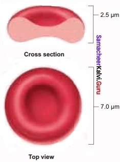
Red blood cells are abundant than other blood cells. There are about 5 million to 5.5 million of RBC mnr3 of blood in a healthy man and 4.5-5.0 million RBC mm ° in healthy women.
The RBCs are very small with a diameter of about 7 pm (micrometer). The structure of RBC is shown in Figure. The red colour of the RBC is due to the presence of a respiratory pigment, hemoglobin dissolved in the cytoplasm.
Hemoglobin plays an important role in the transport of respiratory gases and facilitates the exchange of gases with the fluid outside the cell (tissue fluid). The biconcave-shaped RBCs increase the surface area to volume ratio, hence oxygen diffuses quickly in and out of the cell.
The RBCs are devoid of nucleus, mitochondria, ribosomes and endoplasmic reticulum. The absence of these organelles accommodates more haemoglobin thereby maximising the oxygen-carrying capacity of the cell.
The average life span of RBCs in a healthy individual is about 120 days after which they are destroyed in the spleen (graveyard/cemetery of RBCs) and the iron component returns to the bone marrow for reuse.
Erythropoietin is a hormone secreted by the kidneys in response to low oxygen and helps in the differentiation of stem cells of the bone marrow’ to erythrocytes (erythropoiesis) in adults. The ratio of red blood cells to blood plasma is expressed as Haematocrit (packed cell volume).
White blood cells:
(leucocytes) are colourless, amoeboid, nucleated cells devoid of haemoglobin and other pigments. Approximately 6000 to 8000 per cubic mm of WBCs are seen in the blood of an average healthy individual.
Depending on the presence or absence of granules, WBCs are divided into two types, granulocytes and agranulocytes. Granulocytes are characterised by the presence of granules in the cytoplasm and are differentiated in the bone marrow. The granulocytes include neutrophils, eosinophils and basophils.
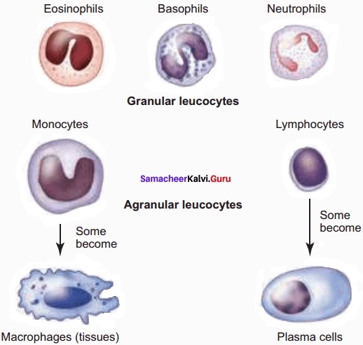
Neutrophils are also called heterophils or polymorphonuclear (cells with 3-4 lobes of nucleus connected with delicate threads) cells which constitute about 60%-65% of the total WBCs. They are phagocytic in nature and appear in large numbers in and around the infected tissues.
Eosinophils have a distinctly bilobed nucleus and the lobes are joined by thin strands. They are non-phagocytic and constitute about 2-3% of the total WBCs. Eosinophils increase during certain types of parasitic infections and allergic reactions.
Basophils are less numerous than any other type of WBCs constituting 0.5%-1.0% of the total number of leucocytes. The cytoplasmic granules are large-sized but fewer than eosinophils.
The nucleus is large-sized and constricted into several lobes but not joined by delicate threads. Basophils secrete substances such as heparin, serotonin, and histamines. They are also involved in inflammatory reactions.
Agranulocvtes are characterized by the absence of granules in the cytoplasm and are differentiated in the lymph glands and spleen. These are of two types, lymphocytes, and monocytes. Lymphocytes constitute 28% of WBCs. These have a large round nucleus and small amount of cytoplasm.
The two types of lymphocytes are B and T cells. Both B and T cells are responsible for the immune responses of the body. B cells produce antibodies to neutralize the harmful effects of foreign substances and T cells are involved in cell-mediated immunity.
Monocytes (Macrophages) are phagocytic cells that are similar to mast cells and have a kidney-shaped nucleus. They constitute 1-3% of the total WBCs. The macrophages of the central nervous system are the ‘microglia’, in the sinusoids of the liver they are called ‘Kupffer cells’, and in the pulmonary region, they are the ‘alveolar macrophages.
Platelets are also called thrombocytes that are produced from megakaryocytes (special cells in bone marrow) and lack nuclei. Blood normally contains 1,50,000 – 3,50,000 platelets mm-3 of blood. They secrete substances involved in the coagulation or clotting of blood. The reduction in platelet number can lead to clotting disorders that result in excessive loss of blood from the body.
![]()
Question 4.
Explain the ABO blood groups?
Answer:
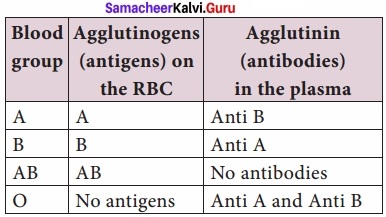
Depending on the presence or absence of surface antigens on the RBCs, blood group in individual belongs to four different types namely, A, B, AB, and O. The plasma of A, B, and O individuals have natural antibodies (agglutinins) in them.
Surface antigens are called agglutinogens. The antibodies (agglutinin) acting on agglutinogen A is called anti-A and the agglutinin acting on agglutinogen B is called anti B.
Agglutinogens are absent in the O blood group. Agglutinogens A and B are present in the AB blood group and do not contain anti-A and anti B in them. A B and O are major allelic genes in ABO systems.
All agglutinogens contain sucrose, D-galactose, N-acetyl glucosamine, and 11 terminal amino acids. The attachments of the terminal amino acids are dependent on the gene products of A and B. The reaction is catalyzed by glycosyltransferase.
Question 5.
Tabulate the distribution of antigens and antibodies in different blood groups?
Answer:
Distribution of antigens and antibodies in different blood groups:
Question 6.
Explain the role of the Rh factor?
Answer:
Rh factor is a protein (D antigen) present on the surface of the red blood cells in the majority (80%) of hum. This protein is similar to the protein present in Rhesus monkey, hence the term Rh. Individuals who carry the antigen D on the surface of the red blood cells are Rh+ (Rh-positive) and the individuals who do not carry antigen D, are Rh– (Rh-negative). Rh factor compatibility is also checked before blood transfusion.
When a pregnant woman is Rh+ and the foetus is Rh+ incompatibility (mismatch) is observed. During the first pregnancy, the Rh antigens of the fetus do not get exposed to the mother’s blood as both their blood are separated by the placenta. However, a small amount of the foetal antigen becomes exposed to the mother’s blood during the birth of the first child.
The mother’s blood starts to synthesize D antibodies. But during subsequent pregnancies, the Rh antibodies from the mother (Rh–) enters the foetal circulation and destroys the foetal RBCs. This becomes fatal to the foetus because the child suffers from anaemia and jaundice. This condition is called erythroblastosis foetalis. This condition can be avoided by administration of anti D antibodies (Rhocum) to the mother immediately after the first childbirth.
![]()
Question 7.
Explain the process of coagulation of blood?
Answer:
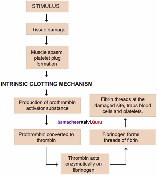
If you cut your finger or when you get yourself hurt, your wound bleeds for some time after which it stops bleeding. This is because the blood clots or coagulates in response to trauma.
The mechanism by which excessive blood loss is prevented by the formation of a clot is called blood coagulation or clotting of blood. The clotting process begins when the endothelium of the blood vessel is damaged and the connective tissue in its wall is exposed to the blood.
Platelets adhere to collagen fibres in the connective tissue and release substances that form the platelet plug which provides emergency protection against blood loss.
Clotting factors released from the clumped platelets or damaged cells mix with clotting factors in the plasma. The protein called prothrombin is converted to its active form called thrombin in the presence of calcium and vitamin K.
Thrombin helps in the conversion of fibrinogen to fibrin threads. The threads of fibers become interlinked into a patch that traps blood cell and seals the injured vessel until the wound is healed.
After sometime fibrin fibrils contract, squeezing out a straw-coloured fluid through a meshwork called serum (Plasma without fibrinogen is called serum). Heparin is an anticoagulant produced in small quantities by mast cells of connective tissue which prevents coagulation in small blood vessels.
Question 8.
Explain the structure of blood vessels?
Answer:
The vessels carrying the blood are of three types; they are the arteries, veins and capillaries. These vessels are hollow structures and have complex walls surrounding the lumen. The blood vessels in hum are composed of three layers, tunica intima, tunica media and tunica externa.
The inner layer, tunica intima or tunica interna supports the vascular endothelium, the middle layer, tunica media is composed of smooth muscles and an extra cellular matrix which contains a protein, elastin. The contraction and relaxation of the smooth muscles results in vasoconstriction and vasodilation. The outer layer, tunica externa or tunica adventitia is composed of collagen fibres.
Arteries:
The blood vessels that carry blood away from the heart are called arteries. The arteries usually lie deep inside the body. The walls of the arteries are thick, non-collapsible to withstand high pressure. Valves are absent and have a narrow lumen. All arteries carry oxygenated blood, except the pulmonary artery.
The largest artery, the aorta (2.5 cm in diameter and 2 mm thick) branch into smaller arteries and culminates into the tissues as feed arteries. In the tissues the arteries branch into arterioles. As blood enters an arteriole it may have a pressure of 85 mm Hg (11.3 KPa) but as it leaves and flows into the capillary, the pressure drops to 35 mm Hg (4.7 KPa). (Note 1 mm Hg = 0.13 KPa.
SI unit of mm Hg is KiloPascal (KPa)). Arterioles are small, narrow, and thin-walled which are connected to the capillaries. A small sphincter lies at the junction between the arterioles and capillaries to regulate the blood supply. Arteries do not always branch into arterioles, they can also form anastomoses.
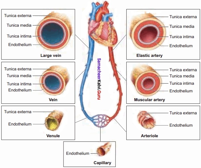
Capillaries:
Capillary beds are made up of fine networks of capillaries. The capillaries are thin-walled and consist of single layer of squamous epithelium. Tunica media and elastin fibres are absent. The capillary beds are the site for an exchange of materials between blood and tissues.
The walls of the capillaries are guarded by semilunar valves. The blood volume in the capillaries is high but the flow of blood is slow. Mixed blood (oxygenated and deoxygenated) is present in the capillaries. The capillary bed may be flooded with blood or maybe completely bypassed depending on the body conditions in a particular organ.
Veins:
Veins have thinner walls and a larger lumen and hence can be easily stretched. They carry deoxygenated blood except, the pulmonary vein. The blood pressure is low and the lumen has a wide wall which is collapsible.
Tunica media is thinner in veins than in arteries. The unidirectional flow of blood in veins is due to the presence of semilunar valves that prevents the backflow of blood. Blood samples are usually taken from the veins rather than artery because of the low pressure in the veins.
![]()
Question 9.
Write a short note on coronary blood vessels?
Answer:
Blood vessels that supply blood to the cardiac muscles with all nutrients and remove wastes are the coronary arteries and veins. Heart muscle is supplied by two arteries namely the right and left coronary arteries.
These arteries are the first branch of the aorta. Arteries usually surround the heart in the manner of a crown, hence called coronary artery (L. Corona – crown). Right ventricle and posterior portion of left ventricle are supplied by the right coronary artery. Anterior and lateral part of the left ventricle is supplied by the left coronary arteries.
Question 10.
Compare the chambers of heart and the methods of circulation in fishes, amphibians, reptiles, crocodiles, birds and mammals?
Answer:
All vertebrates have a muscular chambered heart. Fishes have a two-chambered heart. The heart in fishes consists of sinus venosus, an atrium, one ventricle and bulbus arteriosus or conus arteriosus.
Single circulation is seen in fishes. Amphibian have two auricles and one ventricle and no interventricular septum whereas reptiles except crocodiles have two auricles and one ventricle and an incomplete interventricular septum.
Thus mixing of oxygenated and deoxygenated blood takes place in the ventricles. This type of circulation is called incomplete double circulation. The left atrium receives oxygenated blood and the right atrium receives deoxygenated blood. Pulmonary and systemic circuits are seen in Amphibian and Reptiles.
The Crocodiles, Birds and Mammals have two auricles or atrial chambers and two ventricles, the auricles, and ventricles are separated by inter auricular septum and interventricular septum. Hence there is a complete separation of oxygenated blood from the deoxygenated blood. Pulmonary and systemic circuits are evident. This type of circulation is called complete double circulation.
![]()
Question 11.
Explain the structure of the human heart?
Answer:
The structure of the heart was described by Raymond de Viessens, in 1706. The human heart is made of a special type of muscle called the cardiac muscle. It is situated in the thoracic cavity and its apex portion is slightly tilted towards left. It weighs about 300g in an adult.
The size of our heart is roughly equal to a closed fist. Heart is divided into four chambers, upper two small auricles or atrium and lower two large ventricles.
The walls of the ventricles are thicker than the auricles due to the presence of papillary muscles. The heart wall is made up of three layers, the outer epicardium, middle myocardium and inner endocardium. The space present between the membranes is called pericardial space and is filled with pericardial fluid.
The two auricles are separated by inter auricular septum and the two ventricles are separated by the interventricular septum. The separation of chambers avoids mixing of oxygenated and deoxygenated blood. The auricle communicates with the ventricle through an opening called auriculo ventricular aperture which is guarded by the auriculo ventricular valves.
The opening between the right atrium and the right ventricle is guarded by the tricuspid valve (three flaps or cusps), whereas a bicuspid (two flaps or cusps) or mitral valve guards the opening between the left atrium and left ventricle. The valves of the heart allows the blood to flow only in one direction, i.e., from the atria to the ventricles and from the ventricles to the pulmonary’ artery or the aorta. These valves prevent the backward flow of blood.
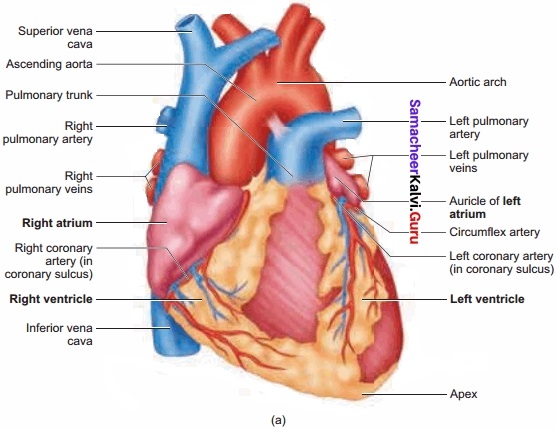
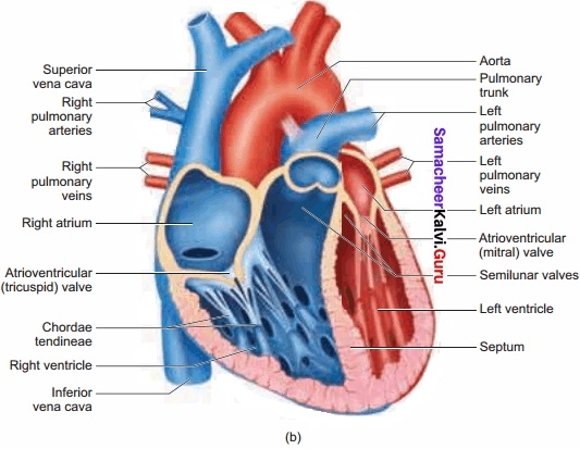
The opening of right and left ventricles into the pulmonary artery and aorta are guarded by aortic and pulmonary valves and are called semilunar valves. Each semilunar valve is made of three half-moon-shaped cusps. The myocardium of the ventricle is thrown into irregular muscular ridges called trabeculae comeae. The trabeculae come are modified into chordae tendinae. The opening and closing of the semilunar valves are achieved by the chordae tendinae.
The chordaetendinae are attached to the lower end of the heart by papillary muscles. The heart receives deoxygenated blood from various parts of the body through the inferior venacava and superior venacava which open into the right auricle. Oxygenated blood from lungs is drained into the left auricle through four pulmonary veins.
![]()
Question 12.
Explain the cardiac cycle?
Answer:
The events that occur at the beginning of the heartbeat and last until the beginning of next beat is called cardiac cycle. It lasts for 0.8 seconds. The series of events that takes place in a cardiac cycle.
PHASE 1:
Ventricular diastole- The pressure in the auricles increases than that of the ventricular pressure. AV valves are open while the semilunar valves are closed. Blood flows from the auricles into the ventricles passively.
PHASE 2:
Atrial systole – The atria contract while the ventricles are still relaxed. The contraction of the auricles pushes the maximum volume of blood to the ventricles until they reach the end-diastolic volume (EDV). EDV is related to the length of the cardiac muscle fibre. The more the muscle is stretched, the greater the EDV and the stroke volume.
PHASE 3:
Ventricular systole (isovolumetric contraction) – The ventricular contraction forces the AV valves to close and increases the pressure inside the ventricles. The blood is then pumped from the ventricles into the aorta without change in the size of the muscle fibre length and ventricular chamber volume (isovolumetric contraction).
PHASE 4:
Ventricular systole (ventricular ejection) – Increased ventricular pressure forces the semilunar valves to open and blood is ejected out of the ventricles without backflow of blood. This point is the end of the systolic volume (ESV).
PHASE 5:
(Ventricular diastole) – The ventricles begin to relax, pressure in the arteries exceeds ventricular pressure, resulting in the closure of the semilunar valves. The heart returns to phase 1 of the cardiac cycle.
![]()
Question 13.
Explain cardiac output in a man?
Answer:
The amount of blood pumped out by each ventricle per minute is called cardiac output (CO). It is a product of heart rate (HR) and stroke volume (SV). Heart rate or pulse is the number of beats per minute. Pulse pressure = systolic pressure – diastolic pressure. Stroke volume (SV) is the volume of blood pumped out by one ventricle with each beat. SV depends on ventricular contraction.
CO = HR x SV. SV represents the difference between EDV (amount of blood that collects in a ventricle during diastole) and ESV (volume of blood remaining in the ventricle after contraction). SV = EDV – ESV. According to Frank-Starling law of the heart, the critical factor controlling SV is the degree to which the cardiac muscle cells are stretched just before they contract.
The most important factor stretching the cardiac muscle is the amount of blood returning to the heart and distending its ventricles, venous return.
During vigorous exercise, SV may double as a result of venous return.
Heart’s pumping action normally maintains a balance between cardiac output and venous return. Because the heart is a double pump, each side can fail independently of the other. If the left side of the heart fails, it results in pulmonary congestion and if the right side fails, it results in peripheral congestion. Frank-Starling effect protects the heart from abnormal increases in blood volume.
![]()
Question 14.
Explain the importance of blood pressure?
Answer:
Blood pressure is the pressure exerted on the surface of blood vessels by the blood. This pressure circulates the blood through arteries, veins and capillaries.
There are two types of pressure, systolic pressure and diastolic pressure. Systolic pressure is the pressure in the arteries as the chambers of the heart contract. Diastolic pressure is the pressure in the arteries when the heart chambers relax.
Blood pressure is measured using a sphygmomanometer (BP apparatus). It is expressed as systolic pressure / diastolic pressure. Normal blood pressure in man is about 120/80mm Hg. Mean arterial pressure is a function of cardiac output and resistance in the arterioles. The primary reflex pathway for homeostatic control of mean arterial pressure is the baroreceptor reflex.
The baroreceptor reflex functions every morning when you get out of bed. When you are lying flat the gravitational force is evenly distributed. When you stand up, gravity causes blood to pool in the lower extremities.
The decrease in blood pressure upon standing is known as orthostatic hypotension. Orthostatic reflex normally triggers baroreceptor reflex. This results in increased cardiac output and increased peripheral resistance which together increase the mean arterial pressure.
![]()
Question 15.
Explain the recording of the electrocardiogram?
Answer:
An electrocardiogram (ECG) records the electrical activity of the heart over a period of time using electrodes placed on the skin, arms, legs, and chest. It records the changes in electrical potential across the heart during one cardiac cycle. The special flap of muscle that initiates the heartbeat is called a sinus-auricular node or SA node in the right atrium.
It spreads as a wave of contraction in the heart. The waves of the ECG are due to depolarization and not due to contraction of the heart. This wave of depolarization occurs before the beginning of the contraction of the cardiac muscle. A normal ECG shows 3 waves designated as P wave, QRS complex and T wave.
P Wave (atrial depolarisation): It is a small upward wave and indicates the depolarization of the atria. This is the time taken for the excitation to spread through atria from SA node. Contraction of both atria lasts for around 0.8-1.0 sec.
PQ Interval (AV node delay): It is the onset of P wave to the onset of QRS complex. This is from the start of depolarisation of the atria to the beginning of ventricular depolarisation. It is the time taken for the impulse to travel from the atria to the ventricles (0.12-0.21 sec). It is the measure of AV conduction time.
QRS Complex: (ventricular depolarisation) No separate wave for atrial depolarization in the ECG is visible. Atrial depolarization occurs simultaneously with ventricular depolarization.
The normal QRS complex lasts for 0.06-0.09 sec. The QRS complex is shorter than the P wave because depolarization spreads through the Purkinje fibers. Prolonged QRS wave indicates delayed conduction through the ventricle, often caused due to ventricular hypertrophy or due to a block in the branches of the bundle of His.
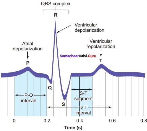
repolarisation. In the heart muscle, the prolonged depolarization is due to retardation of K+ efflux and is responsible for the plateau.
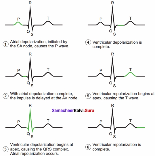
The ST segment lasts for 0.09 sec. T wave (ventricular depolarisation): It represents ventricular depolarisation. The duration of the T wave is longer than QRS complex because repolarisation takes place simultaneously throughout the ventricular depolarisation.
Question 16.
Explain the regulation of cardiac activity?
Answer:
The type of heart in human is myogenic because the heart beat originates from the muscles of the heart. The nervous and endocrine systems work together with paracrine signals (metabolic activity) to influence the diameter of the arterioles and alter the blood flow.
The neuronal control is achieved through autonomic nervous system (sympathetic and parasympathetic). Sympathetic neurons release nor-epinephrine and adrenal medulla releases epinephrine.
The two hormones bind to p – adrenergic receptors and increase the heart rate. The parasympathetic neurons secrete acetylcholine that binds to muscarinic receptors and decreases the heart beat.
Vasopressin and angiotensin II, involved in the regulation of the kidneys, results in vasoconstriction while natriuretic peptide promotes vasodilation. Vagus nerve is a parasympathetic nerve that supplies the atrium especially the SA and the AV nodes.
![]()
Question 17.
What is hypertension?
Answer:
Hypertension is the most common circulatory disease. The normal blood pressure in man is 120/80 mmHg. In cases when the diastolic pressure exceeds 90 mm Hg and the systolic pressure exceeds 150 mm Hg persistently, the condition is called hypertension. Uncontrolled hypertension may damage the heart, brain and kidneys.
Question 18.
Explain the disorder of coronary heart disease?
Answer:
Coronary heart disease occurs when the arteries are lined by atheroma. The build-up of atheroma contains cholesterol, fibres, dead muscle and platelets and is termed Atherosclerosis.
The cholesterol-rich atheroma forms plaques in the inner lining of the arteries making them less elastic and reduces the blood flow. Plaque grows within the artery and tends to form blood clots, forming coronary thrombus. Thrombus in a coronary artery results in a heart attack.
Question 19.
Explain the disorder stroke?
Answer:
Stroke is a condition when the blood vessels in the brain burst (Brain haemorrhage) or, when there is a block in the artery that supplies the brain, (atherosclerosis) or thrombus. The part of the brain tissue that is supplied by this damaged artery dies due to lack of oxygen (cerebral infarction).
Angina pectoris (ischemic pain in the heart muscles) is experienced during the early stages of coronary heart disease. Atheroma may partially block the coronary artery and reduce the blood supply to the heart. As a result, there is tightness or choking with difficulty in breathing. This leads to angina or chest pain. Usually, it lasts for a short duration of time.
![]()
Question 20.
Explain the disorder of myocardial infarction?
Answer:
The prime defect in heart failure is a decrease in cardiac muscle contractility. The Frank-Starling curve shifts downwards and towards the right such that for a given EDV, a failing heart pumps out a smaller stroke volume than a normal healthy heart. When the blood supply to the heart muscle or myocardium is remarkably reduced it leads to death of the muscle fibres.
This condition is called heart attack or myocardial infarction. The blood clot or thrombosis blocks the blood supply to the heart and weakens the muscle fibres. It is also called Ischemic heart disease due to lack of oxygen supply to the heart muscles. If this persists it leads to chest pain or angina. Prolonged angina leads to death of the heart muscle resulting in heart failure.
Question 21.
Explain the disorder of rheumatoid heart disease?
Answer:
Rheumatic fever is an autoimmune disease which occurs 2-4 weeks after throat infection usually a streptococcal infection. The antibodies developed to combat the infection cause damage to the heart. Effects include fibrous nodules on the mitral valve, fibrosis of the connective tissue and accumulation of fluid in the pericardial cavity.
![]()
Question 22.
Explain Cardio Pulmonary Resuscitation (CPR)?
Answer:
In 1956, James Elam and Peter Safar were the first to use mouth-to-mouth resuscitation. CPR is a life-saving procedure that is done at the time of emergency conditions such as when a person’s breath or heartbeat has stopped abruptly in case of drowning, electric shock, or heart attack.
CPR includes rescue of breath, which is achieved by mouth to mouth breathing, to deliver oxygen to the victim’s lungs by external chest compressions which helps to circulate blood to the vital organizer.
CPR must be performed within 4 to 6 minutes after cessation of breath to prevent brain damage or death. Along with CPR, defibrillation is also done. Defibrillation me a brief electric shock is given to the heart to recover the function of the heart.